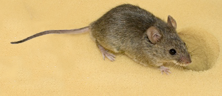
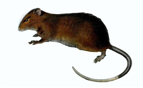
Introduction Mouse Monocytes
The mouse is a species, which is extensively used as a model for the human immune system. Still, when it comes to monocytes the studies are relatively scarce because of the limited amounts of blood available and the difficulty of sampling. Several markers for monocyte identification have been described and subsets of classical, intermediate and non-classical monocytes have been reported. These cells show many similarities to human monocytes but there are also substantial differences including reverse patterns of gene expression (Ingersoll et al, Blood, 115: e10, 2010), such that findings in the mouse cannot be directly applied to man, but need to be confirmed by separate studies on human blood cells.
The first study to clearly demonstrate subsets of mouse monocytes used a genetic model, which employs a GFP-transgene under the control of the CX3CR1-promotor (Palframan et al,J Exp Med,194: 1361, 2001). Here monocytes where defined as F4/80+ cells and a GFPhigh and a GFPlow subset could be identified. In subsequent studies the two subsets were shown to have differential migration patterns (Geissman et al, Immunity, 19: 71, 2003) and the CX3CR1-GFPhigh non-classical monocytes were shown to patrol the vascular endothelium and to transmigrate in response to injury and infection (Auffray et al, Science, 317: 666, 2007). Upon infection with Listeria the non-classical monocytes produced the highest amounts of TNF. Others have used a transgenic model with GFP under the control of the M-CSF-R promotor and with staining for Ly6G to exclude granulocytes. Then, differential staining for GR-1 and for CD43 was employed for separation of subsets. Here the non-classical CD43++ cells were shown to have higher TNF production after stimulation with the TLR4-ligand LPS when compared to classical monocytes (Burke et al, Imm Letters 118: 142, 2008).
The two subset account in average for about 50% each, quite in contrast to man, where the non-classical monocytes at rest make up about 15% of all monocytes.


