
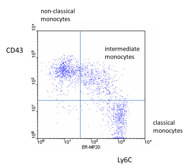
 Dot plot kindly provided by Dr. Pieter JM Leenen, Erasmus Medical Center, Rotterdam, The Netherlands
 Markers and Staining Protocols Mouse Monocytes
In wildtype mice, monocytes have been identified based on low side scatter and high CD11b levels (Sunderkotter et al, J Immunol 172: 4410, 2004) with subsets dissected via differential expression of Ly6C and CD43. Also, monocytes have been defined using low side scatter and staining for CD115 plus F4/80 with GR-1 staining for definition of the subsets (Ingersoll et al, Blood, 115: e10, 2010). A recent paper used light scatter, CD11b and CD115 for monocytes and Ly6C and CD62L for subset definition (Roberts et al, Circulation Research, 126, e61, 2020). In summary, when working with wild type mice, then monocytes can be defined via low side scatter (to exclude granulocytes) and positivity for CD11b, F4/80 and/or CD115, while the subsets can be best identified via differential levels of Ly6C and CD43 or CD62L. The use of two differential markers will ensure that the two population are well separated.
Of note, the use of CD115 for identification of mouse monocytes can be compromised under conditions of severe inflammation, where expression of this cell surface receptor was shown to be downregulated (Drevets et al, J Immunol 185:2432, 2010).